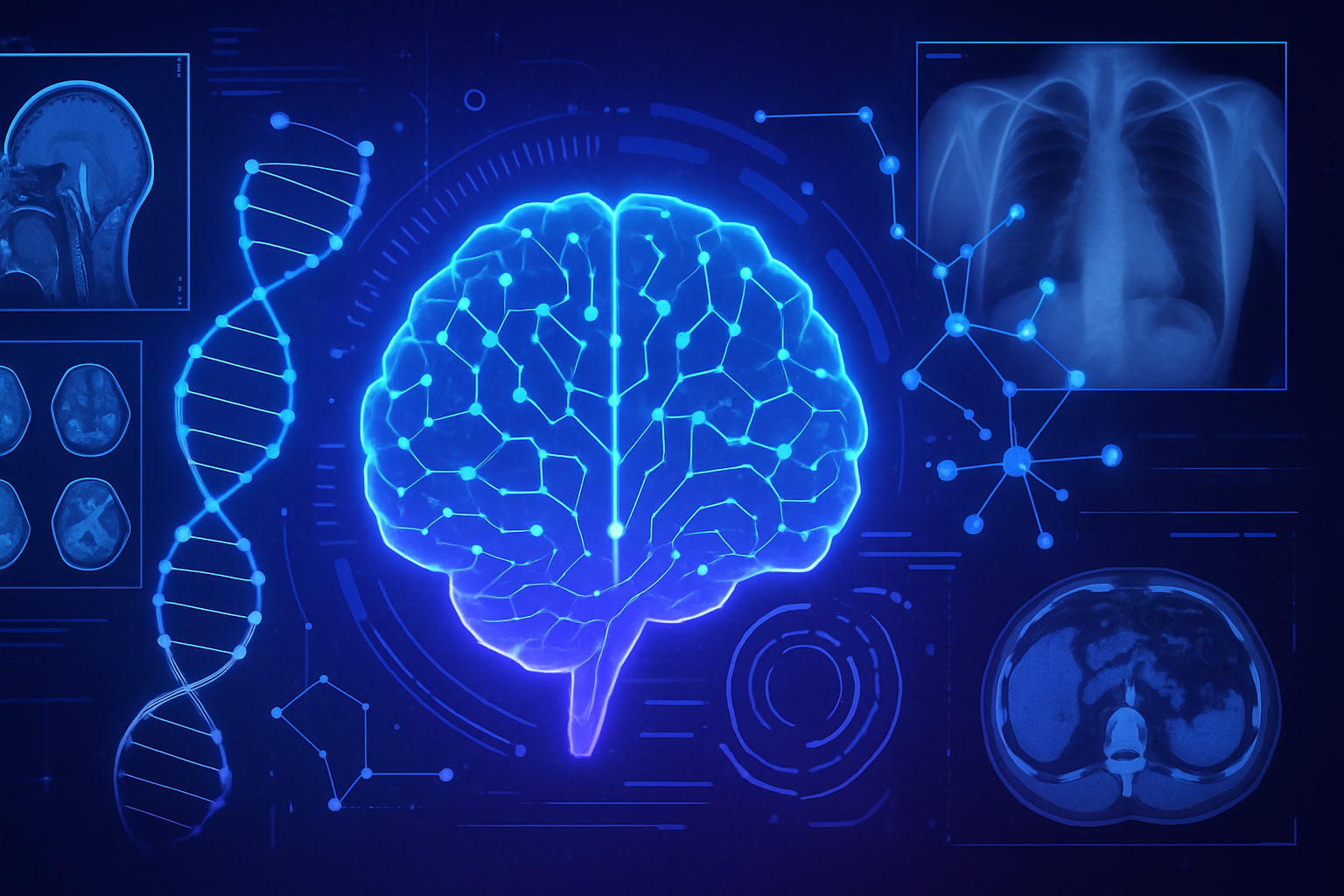
Innovation in artificial intelligence is revolutionizing medical image analysis. A new tool promises to reduce dependence on massive datasets. _Learning from fewer data_ is becoming a reality, facilitating the work of clinicians. This innovative system surpasses traditional methods, making access to accurate diagnostics faster and more cost-effective. _Improvement in image segmentation_ represents a crucial turning point in the fight against various pathologies. This development will transform medical practice in resource-limited settings. _A valuable opportunity for the medical sector_, this new tool could redefine expectations against the limitations of current procedures.
A new AI tool for medical image analysis
An artificial intelligence (AI) tool is revolutionizing medical image analysis, thereby facilitating the development of dedicated software for doctors and researchers. This innovative mechanism proves particularly effective when patient scan samples are limited, posing a major challenge in the medical field.
Optimization of medical image segmentation
This new tool improves the process of medical image segmentation, a technique where each pixel is labeled based on its content, for example, cancerous or healthy tissue. Traditionally, this work requires the intervention of highly qualified specialists, making the process costly and time-consuming.
Research conducted by Li Zhang, a PhD student at the University of California, San Diego, highlights the limitations of deep learning methods, which are traditionally data-hungry. The models require a vast amount of annotated images to learn effectively. This insatiable need for annotated data often hinders advancements in the field.
Reduced data requirements
Thanks to the innovation from Zhang and his colleagues, a novel process allows AI to learn from a limited number of samples labeled by experts, thus reducing the data requirement by a factor of up to 20. This advancement could make the creation of diagnostic tools more accessible and cost-effective, particularly in healthcare facilities with limited resources.
In tests, this AI tool showed a 10 to 20% improvement in model performance in situations where annotated data was scarce. Impressive results included identifying skin lesions in dermoscopy images and detecting breast cancer in ultrasounds.
Functioning of the AI system
The system operates in several steps. Initially, it learns to generate synthetic images from segmentation masks, which are color overlays indicating corresponding parts of an image. Subsequently, this knowledge is utilized to create new artificial image-mask pairs, thus augmenting a small sample of real data.
A segmentation model is then trained using this new data. The continuous feedback loop plays a fundamental role in the tool’s effectiveness. It helps guide data generation while considering the model’s learning quality. The synthetic data is therefore not only realistic but also specifically tailored to enhance the model’s segmentation capabilities.
Future perspectives and clinical applications
The team envisions increasing the intelligence and versatility of this tool. A key point lies in integrating clinician feedback directly into the training process. This targeting could enhance the relevance of generated data for real medical applications.
A promising future emerges, where this type of tool could significantly simplify disease identification through medical images, particularly in dermatology. For example, a dermatologist might only need to annotate 40 images to help the AI detect suspicious lesions in real-time.
This discovery paves the way for faster and more accurate diagnostics while making medical image analysis tools much more accessible.
To learn more about other advancements related to AI, you can refer to articles such as those on optimizing neocortex calculations or on the revolutionary impact of AI in ophthalmology.
Frequently asked questions about the AI tool for medical image analysis
What is this AI tool for medical image analysis?
It is an innovative technology that uses artificial intelligence to perform segmentation of medical images, thereby classifying pixels based on what they represent, such as normal or cancerous tissues.
How does this AI tool improve image analysis with less data?
It can learn to segment images with a reduced number of samples annotated by experts, requiring up to 20 times less data compared to traditional methods.
What types of medical images can be analyzed with this tool?
The tool has been tested on various types of images, including dermoscopy to identify skin lesions, ultrasounds to detect breast cancer, fetoscopic images to visualize placental vessels, and standard photos for foot ulcers.
What is the impact of this tool on medical diagnosis?
This tool can assist doctors in making quicker and more accurate diagnoses, reducing the need for manual annotations of thousands of images to just a few dozens.
How does the tool generate synthetic data?
It learns to create artificial images from segmentation masks, thus augmenting a small set of real data and improving model performance through a continuous feedback loop process.
Is this technology already used in clinics?
Although it is still in the research phase, the potential of this technology is promising for future integration into clinical environments, particularly where resources are limited.
What are the benefits of this approach compared to traditional methods?
Doctors and researchers can benefit from a less costly and faster training of medical imaging software while achieving segmentation performance comparable to, or even exceeding, that of standard methods.
How will the tool evolve in the future?
Researchers plan to enhance the intelligence of the tool and adapt it by directly incorporating clinician feedback to make the generated data more relevant for real medical use.


